|
||
|
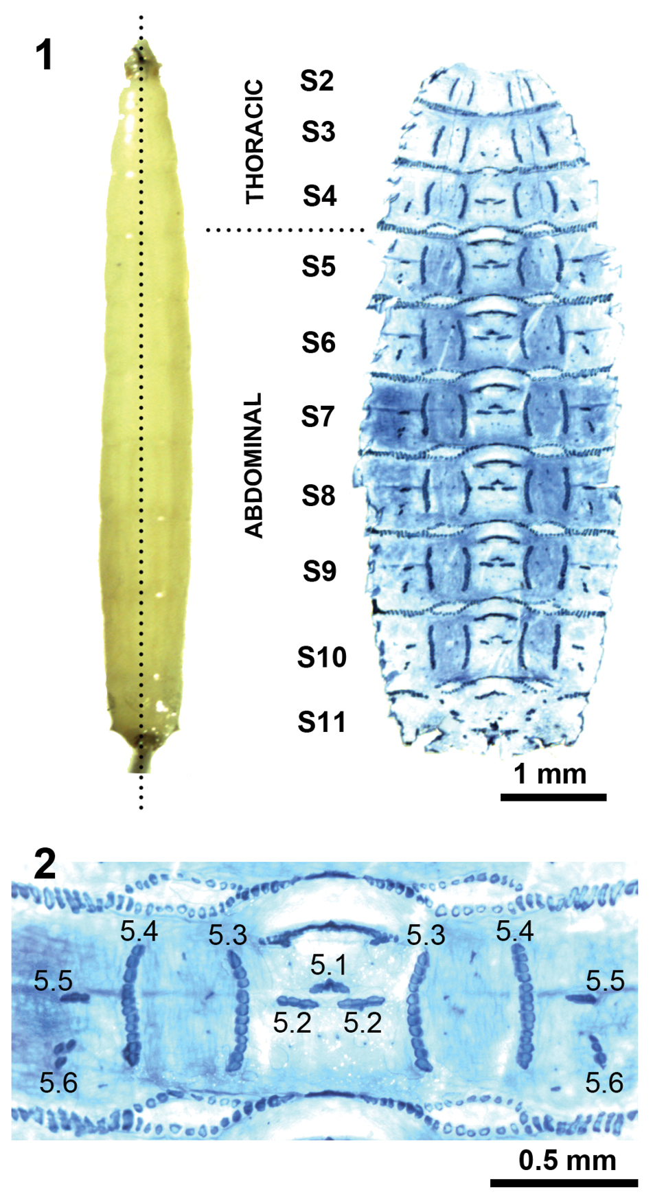
Liopiophila varipes (Meigen), third-instar larva. 1. Dorsal view of a pinned larva before dissection (left) and stained larval cuticle (right), showing the symmetrical muscular attachment sites on segments S2 to S11; 2. Detail of the abdominal segment S5 showing the label for each muscular attachment site pattern. |