|
||
|
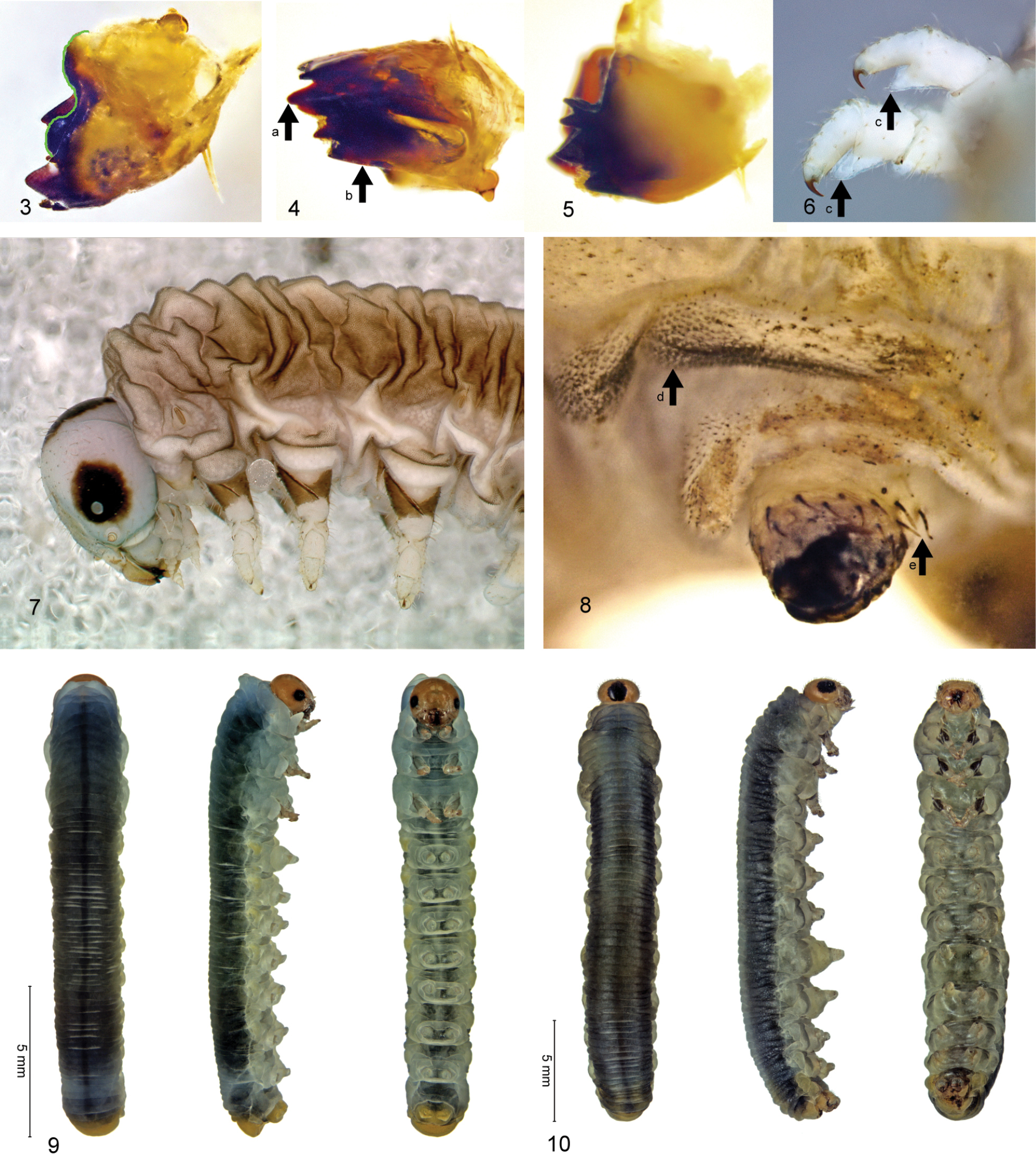
Xenapates larvae: 3. X. braunsi left mandible, outer face; green line along edge of mesal ridge. 4. X. braunsi left mandible, ventral; (a) medial tooth, (b) inner face. 5. X. braunsi right mandible, inner face. 6. X. gaullei right pro- and mesothoracic legs; (c) expanded apex of femur. 7. X. gaullei thorax, lateral. 8. X. braunsi proleg and ventral part of abdominal segment 8, external surface; (d) reticulate-spiculate surface structure on surpedal lobe; (e) setae on proleg (some missing). 9. X. braunsi mature larva; dorsal, lateral and ventral (from left). 10. X. gaullei mature larva. |