|
||
|
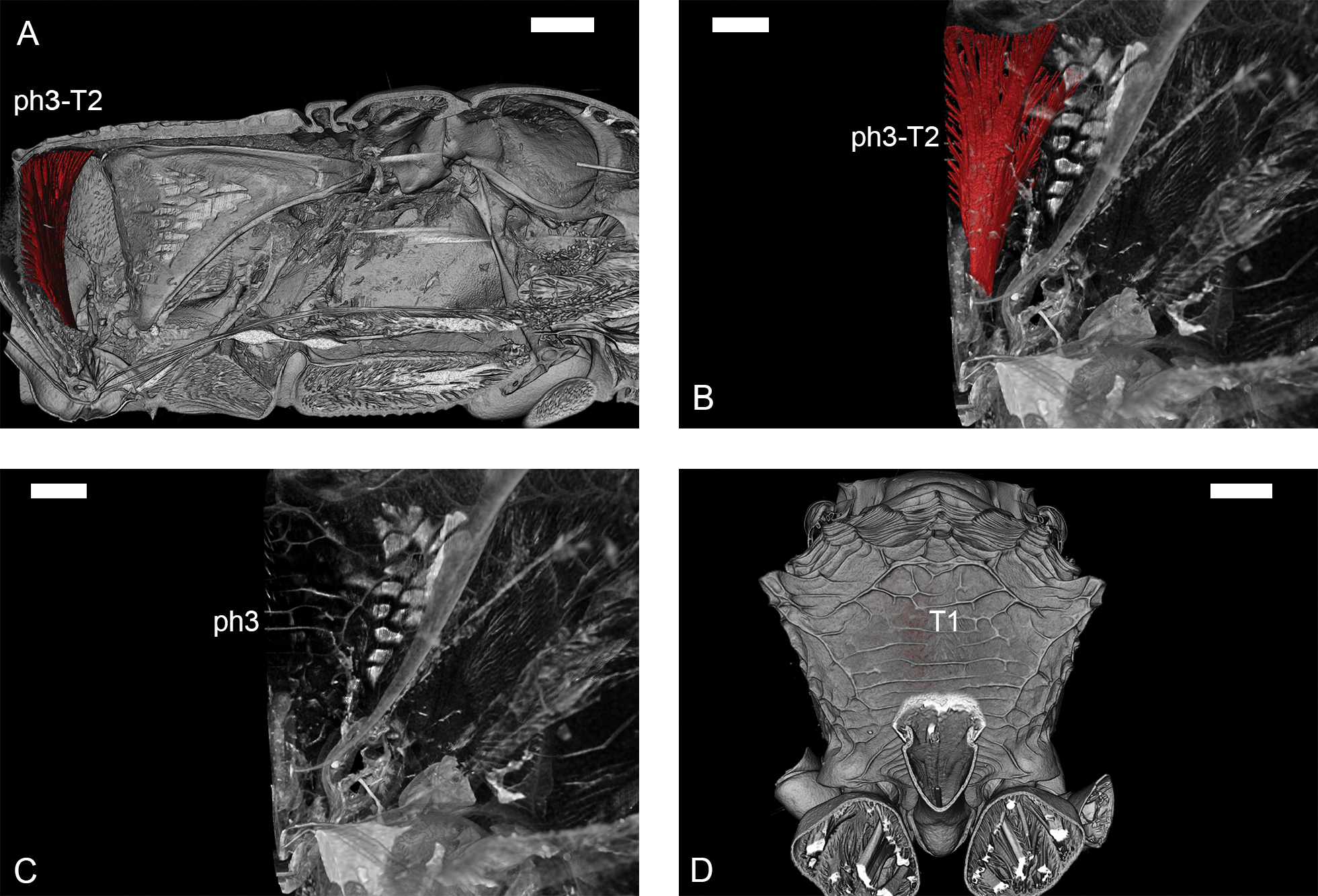
Illustration of the metaphragma (ph3) in the propodeum (T1) of Ampulex compressa. A. Medial view, anterior to the right; B. Anteromedial view on ph3-T2 – metaphragmo-second abdominal tergal muscle; C. Anteromedial view on ph3; D. Posterior view on the vertical part of propodeum. Scale bars: 0.6 mm (A), 0.3 mm (B, C), 0.9 mm (D). |