|
||
|
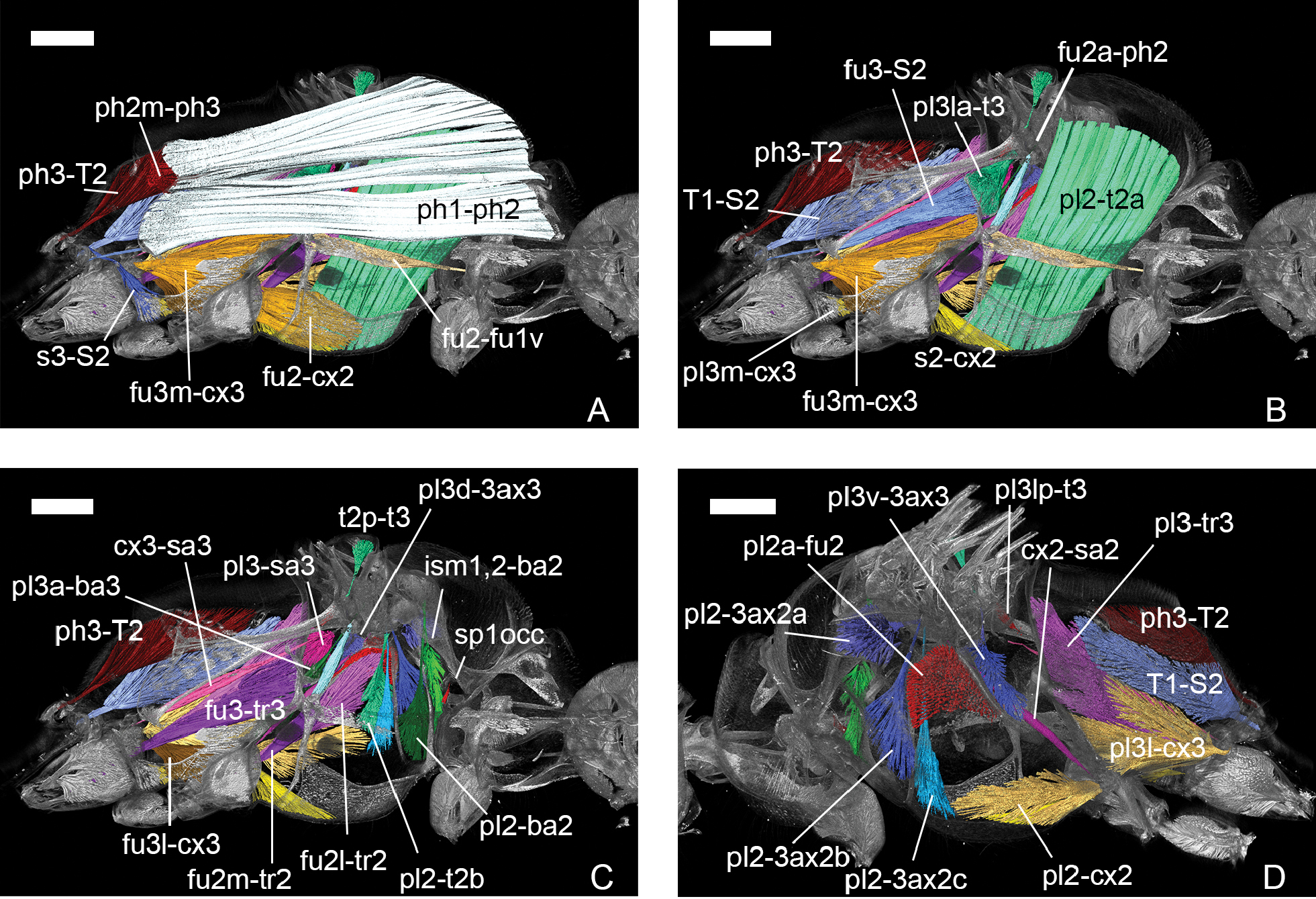
Sceliphron destillatorium, volume rendering, mesosomal musculature, A–C: medial view, anterior to the right, D: lateral view, anterior to the left. A. Muscles discernible from the centre; B. Muscles positioned sublateral; C. Muscles located sublateral and lateral; D. Laterally positioned muscles. Abbreviations: fu2-fu1v – ventral mesofurco-profurcal; pl2-t2a – first mesopleuro-mesonotal; pl2-ba2 – mesopleuro-mesobasalar; sp1occ – anterior thoracic spiracle occlusor; ism1,2-ba2 – intersegmental membrane-mesobasalar; pl2-3ax2a – first mesopleuro-third axillary sclerite of fore wing; pl2-3ax2b – second mesopleuro-third axillary sclerite of fore wing; pl2-3ax2c – third mesopleuro-third axillary sclerite of fore wing; pl2-t2b – second mesopleuro-mesonotal; cx2-sa2 – mesocoxo-mesosubalar; fu2a-ph2 – anterior mesofurco-mesolaterophragmal; pl2a-fu2 – anterior mesopleuro-mesofurcal; pl2-cx2 – mesopleuro-mesocoxal; s2-cx2 – mesosterno-mesocoxal; fu2-cx2 – mesofurco-mesocoxal; fu2l-tr2 – lateral mesofurco-mesotrochanteral; fu2m-tr2 – median mesofurco-mesotrochanteral; ph1-ph2 – prophragmo-mesophragmal; pl3a-ba3 – anterior metapleuro-metabasalar; t2p-t3 – posterior mesonoto-metanotal; pl3la-t3 – anterolateral metapleuro-metanotal; pl3d-3ax3 – dorsal metapleuro-third axillary sclerite of hind wing; pl3-sa3 – metapleuro-metasubalar; cx3-sa3 – metacoxo-metasubalar; pl3m-cx3 – median metapleuro-metacoxal; fu3l-cx3 – lateral metafurco-metacoxal; fu3m-cx3 – median metafurco-metacoxal; pl3l-cx3 – lateral metapleuro-metacoxal; fu3-tr3 – metafurco-metatrochanteral; pl3-tr3 – metapleuro-metatrochanteral; ph2m-ph3 – median mesophragmo-metaphragmal; ph3-T2 – metaphragmo-second abdominal tergal; T1-S2 – propodeo-second abdominal sternal; fu3-S2 – metafurco-second abdominal sternal; s3-S2 – metasterno-second abdominal sternal. Scale bars: 0.8 mm (A–C), 0.9 mm (D). |