|
||
|
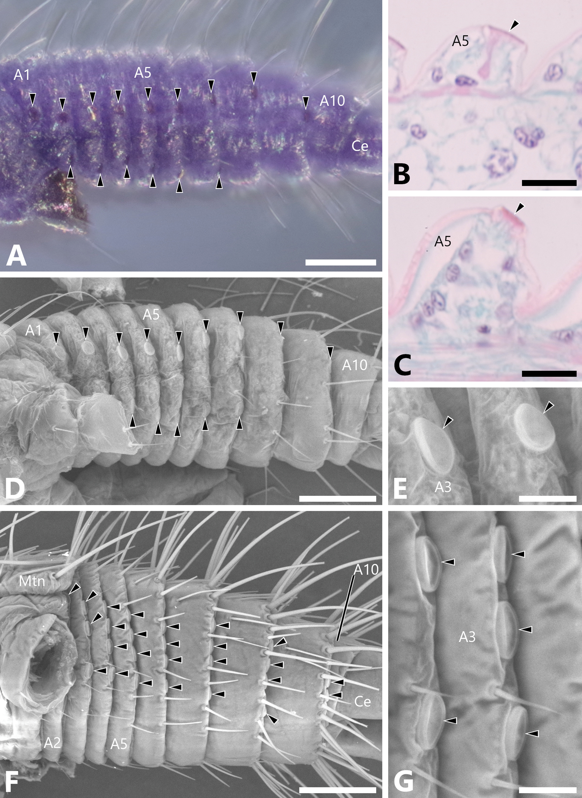
Chloride cells of first instar nymphs of Microperla brevicauda and Yoraperla uenoi, anterior to the left. A. Abdomen of M. brevicauda, lateral view, stained with Mayer’s acid haemalum; B, C. Horizontal sections of fifth abdominal segment of M. brevicauda (B) and Y. uenoi (C); D, E. Abdomen of M. brevicauda, lateral view, scanning electron microscopy (SEM), all abdominal segments (D) and enlargement of chloride cells (E); F, G. Abdomen of Y. uenoi, ventrolateral view, SEM, all abdominal segments (F) and enlargement of chloride cells (G). Arrowheads show the chloride cells. Abbreviations: A1, 2, 3, 5, and 10: first, second, third, fifth and tenth abdominal segments, respectively; Ce, cercus; Mtn, metanotum. Scale bars: 50 µm (A, D, F); 10 µm (B, C, E, G). |