|
||
|
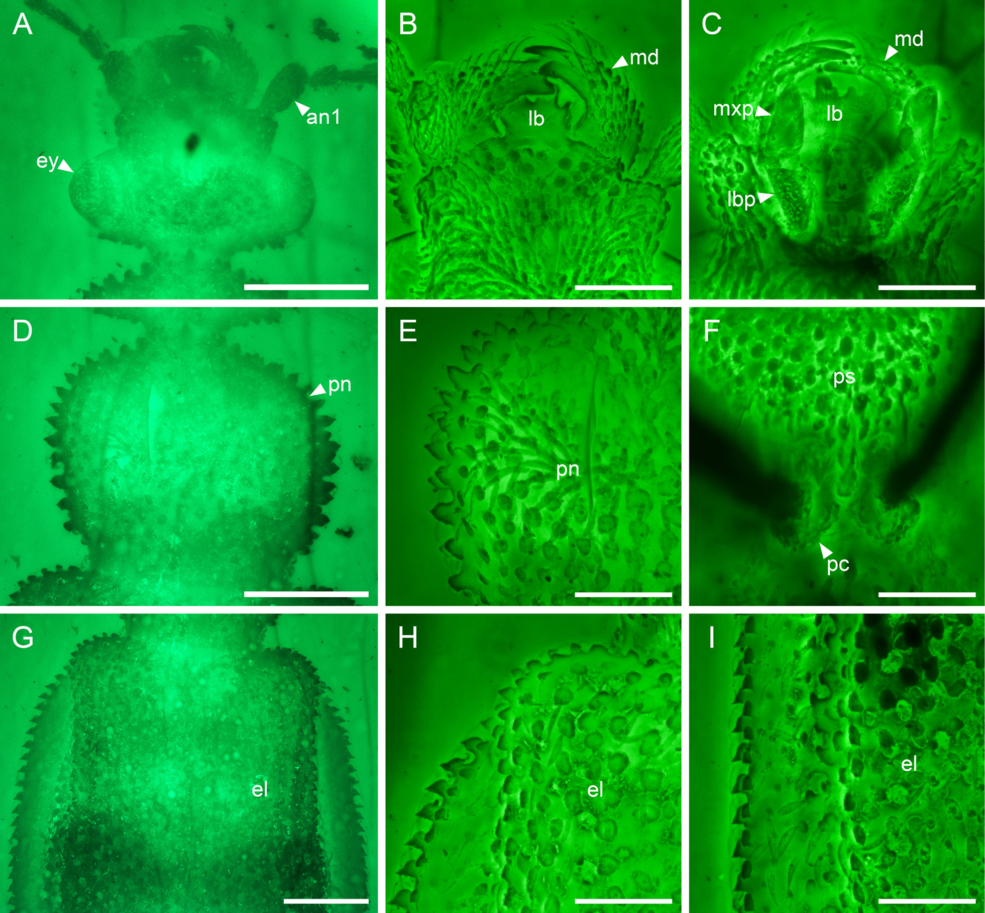
Details of Omma cf. manukyani, NIGP176635, under widefield fluorescence (A, D, G) or confocal microscopy (B, C, E, F, H, I), with the relative positions of the confocal images labeled in Suppl. material 1. A–C. Head, dorsal view (A, B) or ventral view (C); D–F. Prothorax, dorsal view (D, E) or ventral view (F); G–I. Elytra, dorsal view. Abbreviations: an1, antennomere 1; el, elytron; ey, compound eye; lb, labrum; lbp, labial palp; md, mandible; mxp, maxillary palp; pc, procoxa; pn, pronotum; ps, prosternum; v4,5, ventrites 4,5. Scale bars: 600 μm (A, D, G); 300 μm (B, C, E, F, H, I). |