|
||
|
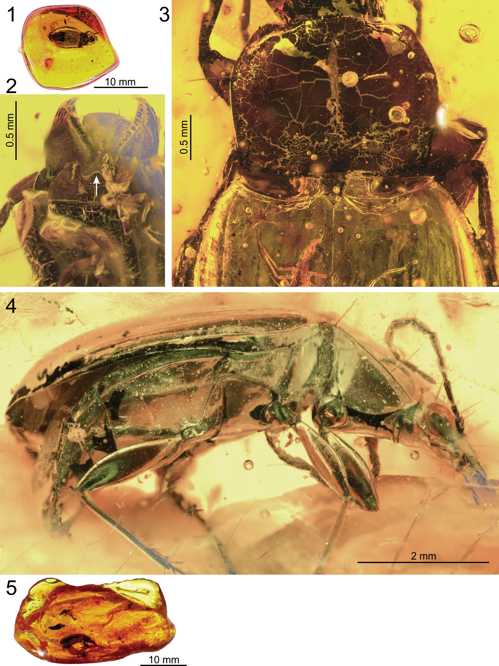
Quasicalathus elpis (Ortuño and Arillo 2009), light microscopic images of specimens “Groehn 4879” (1–3.) and “Groehn 7814” (4, 5.). 1, 5. General view of the amber pieces; 2. Ventral side of head (the white arrow points to the mentum tooth; note that the mentum is somewhat detached from the head capsule); 3. Pronotum and anterior part of elytra showing the markedly concave basal margin and projected humeri; 4. Right lateral view of body. |