|
||
|
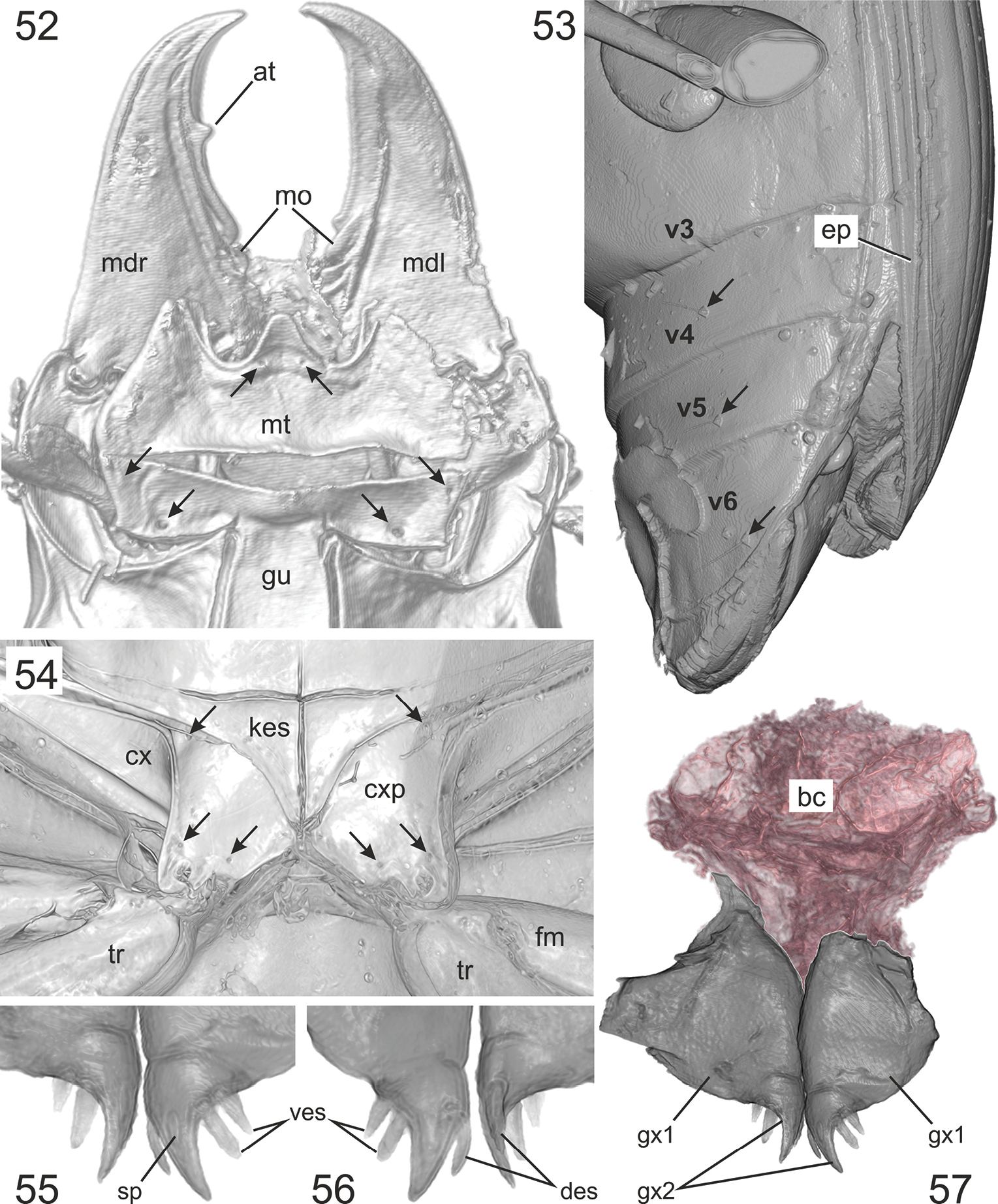
Quasicalathus elpis (Ortuño and Arillo 2009), volume rendering of specimen “Groehn 7889”. 52. Head, ventral aspect (the arrows point to the insertions of the setae near base of mentum tooth and on submentum); 53. Abdomen, left lateral aspect (the arrows point to the insertions of the setae on ventrites IV, V, and VI); 54. Metacoxal area (the arrows point to the insertions of the coxal setae); 55. Apical gonocoxites, ventral aspect; 56. Apical gonocoxites, dorsal aspect; 57. Gonocoxites and remains of the bursa copulatrix (the latter was highlighted by red colour using the segmentation function of Amira software). Abbreviations: at – apical tooth of retinacle; kes – metathoracic katepisternum; cx – metacoxa; cxp – metacoxal plate; des – dorsal ensiform setae; ep – elytral epipleuron; fm – metafemur; gu – gula; mdl – left mandible; gx1 – basal gonocoxite; gx2 – apical gonocoxite; mdr – right mandible; mo – molar; mt – mentum; sp – sensory pit; tr – metatrochanter; ves – ventral ensiform setae; v3, v4, v5, v6 – ventritres III, IV, V, VI. |